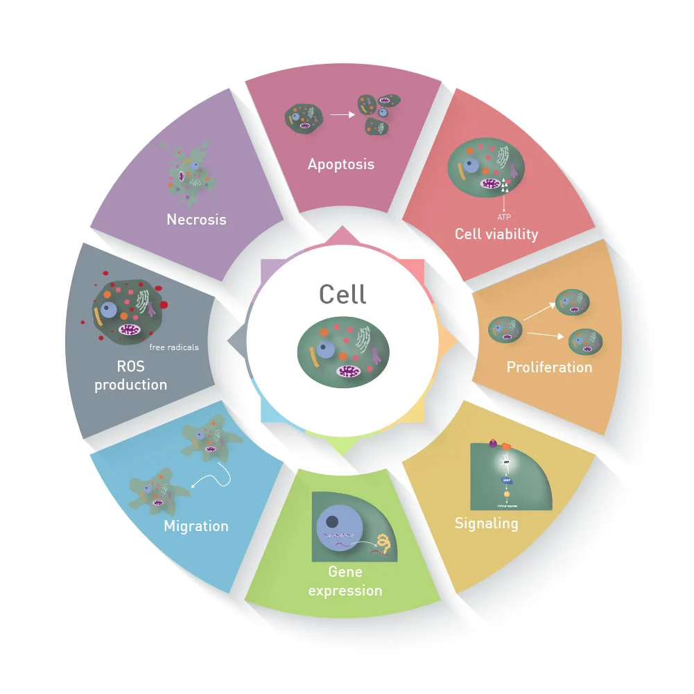
Flow cytometry is a crucial component of bioanalytical laboratories. Flow cytometry cell-based assays employ a laser-based system to detect and evaluate physical and chemical properties of cells or particles. Flow cytometry has multiple applications through different assay formats, such as cell-based functional assays and cell-based screening assays. Each assay format has a unique feature applicable to distinct techniques.
Flow cytometry assessments follow the principle of gating. Regions and gates are marked around the cell population with similar characteristics, such as marker expression, forward scatter, and side scatter expressions, to assess and quantify the cell population of interest. The current article discusses gating strategies and data analysis for flow cytometry in cell-based assays.
Gating strategies and data analysis
Comprehending intricate details about experimental controls and the target cell population is critical before beginning flow cytometry experiments. This information could include simple elements such as cell size and their ability to change during the experiment. Such data is crucial, especially for intracellular staining, as the ligation and fixation process can change the granularity and cell size, resulting in an altered side and forward scatter profile. In the case of cells having known markers, including them during the staining process will aid in determining the cells of interest. On the other hand, having a known negative will allow us to establish negative gates and identify the real cell population.
The primary step in gating is to differentiate the cell population based on their side and forward scatter properties. Forward scatter helps estimate the size of the cell population, whereas side scatter gives a value of the granularity of the cell population. However, these values depend on multiple factors such as the sheath fluid, laser wavelength, study sample, and the sample refractive index and collection angle.
Distinguishing the cell population can be relatively simple for cell lines with a single cell type. However, the complexity increases for samples containing multiple cell types. For example, red-cell lysed whole blood contains different cell populations, resulting in blue, green, yellow, and red hotspots for each type of cell population. The light scatter plot for lymphocytes, monocytes, and granulocytes can help them distinguish from dead cells and cellular debris. Often, dead cells and cellular debris have a lower forward scatter and hence, are located at the bottom left corner of the density plot. However, scientists can increase the forward scatter threshold to avoid these data points or discard them by gating the cell population of interest. This gating strategy is ideal to remove dead cells with increased nonspecific binding of antibodies and autofluorescence. Additionally, researchers can use a viability dye, which can be more reliable and effective.
The cells or events inside a gate can then be analyzed for marker expression. Researchers can express the data using histograms. However, they should conduct flow cytometry experiments using appropriate controls, such as unstained controls and isotypes, for accurately identifying positive data sets. Graph formats such as two-parameter density plots can also be employed in flow cytometry evaluations for cell-based screening assays.


